
Procedure
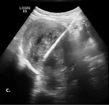
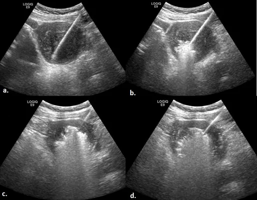
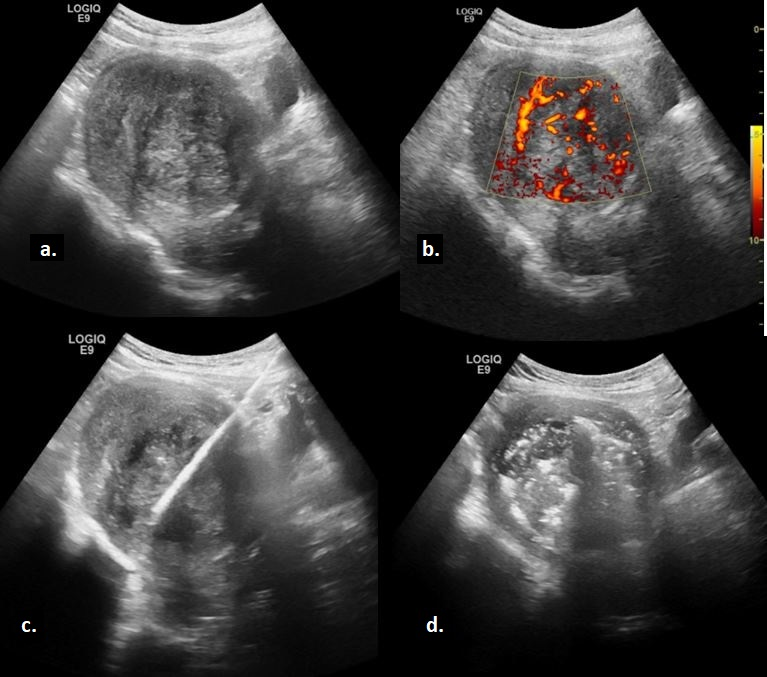
The ablations were performed under general anesthesia through intravenous injection of propofol (0.5–2.0 mg/kg/h), fentanyl (1–2 μg/kg), and midazolam (0.07–0.08 mg/kg). An anesthesiologist was present during the entire procedure. A local anesthetic (10 mL solution of lidocaine 2%) was injected at the entrance site of the antenna. All patients received antibiotic prophylaxis with a preprocedural intravenous injection of 1 g of cefazolin. Patients were placed in a supine position for the treatment. An interventional radiologist (S.A.) with 25 years of experience (10 years of experience in ablation) inserted the microwave antenna into the targeted fibroid using the most suitable US probe (transabdominal for anterior wall fibroid/anteverted uterus or transvaginal for posterior wall fibroid/retroverted uterus). The output energy of 100 W for a total time between 8 and 20 minutes was set to induce ablation. The entire ablation process was monitored via real-time ultrasonography (Fig 1c). MWA was stopped when the hyperechogenic signal covered the entire lesion or extended to 3–5 mm from the margin of the serosa or uterine endometrium (Fig 1d). If the fibroid exceeded 5 cm in diameter, the antenna was partially withdrawn and repositioned to treat the entire mass. The patients were observed in the clinic overnight to monitor for any acute AEs.
Indications
Contradications
Adverse Events
- fibroid diameter of ≥4 cm and <10 cm
- absence of perimenopausal signs
- FIGO Type 7
- cervical, ovarian, uterine, or endometrial malignancy
- severe and uncorrectable coagulopathy
- uterine perforation
- pelvic organ injury